
Folliculometry – The best way for Ovulation Tracking.
Ovulation is a crucial event in the menstrual cycle of women. Ovulatory disorders are one of the main reasons of infertility in women. Before the introduction of ultrasound monitoring, doctors depended on the signs & symptom of the ovulation which were not always conclusive.
Hackeloer, in 1978, first introduced that the developing graffian follicle in the ovary can be monitored in the normal menstrual cycle. Then this technique has been adopted rapidly to monitor follicular growth in both natural and stimulated cycles as well. Since then ultrasonography became the diagnostic tool in investigating follicular development process and ovulation.
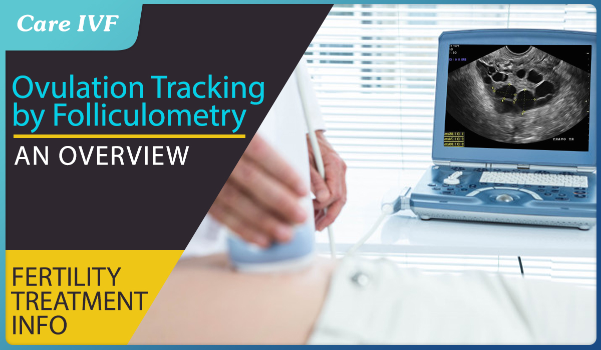
Subsequently, Transvaginal sonography (TVS), was introduced which allows visualisation of finer ovarian details.
The major advantages of transvaginal ultrasound scanning (TVS) include:
- A more accurate localisation and visualisation of the ovary
- Allowing early follicles to be examined and followed throughout the cycle.
- It can measure small follicle of 2-3mm.
- Full bladder is not necessary with TVS and hence the procedure is more comfortable, easy to schedule technique.
- High frequency probe used in TVS provides better axial and lateral resolution
WHAT IS FOLLICULOMETRY?
It is a series of ultrasonography mainly done in transvaginal route starting from day 2 of the menstrual cycle upto documentation of rupture of follicle. It is randomly used in natural cycle, IUI cycle and IVF Cycle. It can be devided into 3 phases. It exactly follows the physiological growth of follicle in follicular phase.
- Baseline scan
- Preovulatory scan
- Luteal phase scan
FOLLICULAR PHASE ASSESSMENT VIA ULTRASOUND SCANS:
BASELINE SCAN
When it’s done - done in day 2 or day 3 of menstrual cycle.
Rational: In the start of menstrual cycle, the Ovaries are silent (in resting phase) and the hormonal level are at baseline. It is easier to see the anatomical details of the ovary and adnexa (the appendages of the uterus, namely the ovaries, the Fallopian tubes, and the ligaments that hold the uterus in place) along with endometrium. We can increase the accuracy by USG with Colour Doppler, 3D USG and Power Doppler.
Baseline scan helps to assess the ovarian response and ovarian reserve.
What is Ovarian Response?
Ovarian response is the how the ovaries respond to stimulation medication and is very important for planning an IUI or IVF cycle. It can help identify women who respond poorly to a stimulation medications and thus help plan the dosage for better response.
What is Ovarian Reserve?
Women all born with all the eggs they can ever produce. Eggs are formed from primordial follicles and the ovaries act as an ovarian bank/ reserve for these primordial follicles. Ovarian Reserve is the number of eggs that remain this ovarian pool and is a major determinant of a woman’s fertility.
BASELINE SCAN APLICATIONS:
- Measure the Ovarian size and volume –
The volume of the ovaries can be correlated with ovarian response. The volume of each ovary is calculated using the formula (Ovary length x Ovary width x Ovary height x 0.5). The Normal ovarian volume of both ovaries combined is 10 ml.
- Women with small ovaries (volume of less than 4 ml) have a poor ovarian response.
- Ovarian volume <3cc predict poor response of ovary to any controlled ovarian hyper stimulation.
- Can differentiate between polycystic/normal sized ovaries.
- Check the Ovarian Position - whether ovary is in proper anatomical place or in displaced location (high up/attached to posterior surface of uterus/ Peri-ovarian adhesion - can assess regarding endometriosis or PID by seeing adhesion.)
- Check presence of any cyst / pathology of Adnexa
- Functional cycts, Endometriotic cyst, dermoid cyst, Haemorrhagic cyst Paraovarian cyst (cyst completely separate from ovarian structure), fimbrial cyst
- Adnexal pathology – problems in the tubes, hydrosalpinx, pyosalpinx e.t.c
- ANTRAL FOLLICLE COUNT/ N.P.O (follicle number per ovary) AFC is a predictor of Ovarian Reserve (the no of remaining primordial follicles or egg precursors a women has in her ovaries). Research shows that the size of the pool of Antral follicles is heavily influenced by the size of the pool of remaining primordial follicles from which they were recruited. That is the number of AFC recruited in every cycle is proportionate to the number of remaining egg forming cells in the ovarian reserve. Therefore in every cycle, by counting the F.N.P.O, it’s possible to predict the how big or how small the size of the ovarian reserve is. In Assisted Reproductive cycles, AFC is a major determinant of follicular response to any stimulation drug.
a. If it is <4 in both ovaries then it is a poor response predictor and if more than 16 then it is going to give higher response (NICE).
b. The ovarian response to any drug can be more accurate if we combine the F.N.P.O with values of Anti Mullerian Hormone (AMH) and Basal F.S.H by giving blood for testing on D2/3.
c. There Linear correlation seen between AFC and AMH. Which means higher the AFC higher the AMH and AFC is low, AMH will be low.
Advantage of AFC
It shows the best correlation with women’s age and declines linearly 3.8% per year according to Ng et al & it decrease 4.8% before 37 years and 11.7% after 37 years.
- Doing AFC alone is more cost effective for predicting ovarian response.
- When we combine AFC and stromal blood flow (visualised via USG colour Doppler) – it allows a more precise prediction of ovarian response in IVF cycle.
- Stromal flow index:
- <11 –low responder,
- 11-14-good,
- >15-risk of Ovarian Hyperstimulation.
Clinical application - In stimulated cycle, if we combine AFC, age, ovarian volume and a few other USG parameters it helps us in the dose calculation of gonadotropins in ART cycles.
PRE-OVULATORY SCAN:
It involves measuring the growing follicle via a series of scans done during the follicular phase of the menstrual cycle and starts from:
- Day 9 or 10 of natural or IUI cycle
- Day 5 or 6 of IVF cycle and serial scan
It basically tracks the physiological growing of the follicle throughout the follicular phase so that the ovulation can be predicted.
Purpose of serial scans -
- To evaluate Ovarian Response to Stimulation.
- Monitor the effect of Pituitary Down Regulation
- Avoid Ovarian Hyper Stimulation Syndrome OHSS
- Identify and measure the Growing follicle Which becomes dominant follicle (measurement taken when a follicle is seen as a rounded structure
- is more than 10mm in diameter
- grows at a rate of 2-3mm /day
- has no internal echogenicity
- has thin regular wall
- Measure the Mature follicle (at around day 13-14 in a regular cycle)
- is 16-18mm,
- has thin wall ,
- Surrounded by thin hypo-echoic rim (30-40%),
- Sometimes a cumulus structure can be seen. This cumulus structure develop 36 hours prior to rupture. A low level echo can be seen in follicle parallel to the wall.
- To decide the correct timing of administering the hCG injection for IUI /Ovum pick up in IVF cycle by monitoring changes in the dominant follicle as it matures and checking for all the other optimal features like increase in vascularity. If all above optimum features of follicles are present then it’s good enough for trigger.
MATURE DOMINANT FOLLICLE IN 2D AND COLOR DOPLER
- In stimulated cycles (IUI / IVF) it helps check if the ovulation stimulation is optimum, whether the parameters suggest a cycle to be cancelled, and also decide the timing for trigger.
- Monitor Endometrium Response In Follicular Phase
Like the ovarian follicles, the endometrium as well undergoes a series of physiological changes in preparetion for implantation should the egg be fertilized.
Endometrium growth is divided into four zones and each of these phases have distinctive physiology that can be detetced in USG using Color / Power doppler.
Measuring the endometrial growth has a major clinical application in treatment outcome
- As the Pregnancy rate is found to increase proportionately from zone 1 to zone 4 from 7.55% to >40% in zone
- No pregnancy rate with endometrial thickness <3 and volume <3 -7 cc and better pregnancy rate with endometrial thickness > 7mm and volume >7cc
- Absence of blood flow in endometrial and sub endometrial zone on day of hCG injection indicate failure of implantation.
LUTEAL SECRETORY PHASE ASSESSMENT
Rupture of follicle leads to release of egg and then it collapses and forms corpus lutein and causes the release of progesterone. Progesterone is responsible for the endometrial preparation.
With this scan we can establish the presence of an adequately functioning corpus luteum. It will show luteal blood flow in Colour Doppler with R.I 0.35-0.50 and PSV10-15. The endometrium becomes hyper echoic and outer margin becomes fluffy and blurred.
Clinical significance: Helps confirm the final physiological changes in ovaries and uterus.
FOLLICULOMETRY IN A NUT SHELL
- Folliculometry is an excellent tool for assessment of menstrual cycle and tracking the follicular growth till ovulation
- Helps to interpret hormonal synchrony of menstrual cycle
- It helps predict the date of ovulation
- Only modality for surest documentation of ovulation and very cost effective, becomes the most essential tool for any ART procedure
- TVS gives much superior accurate diagnosis of follicular monitoring
Article Tags
About the author
Leave a Comment