
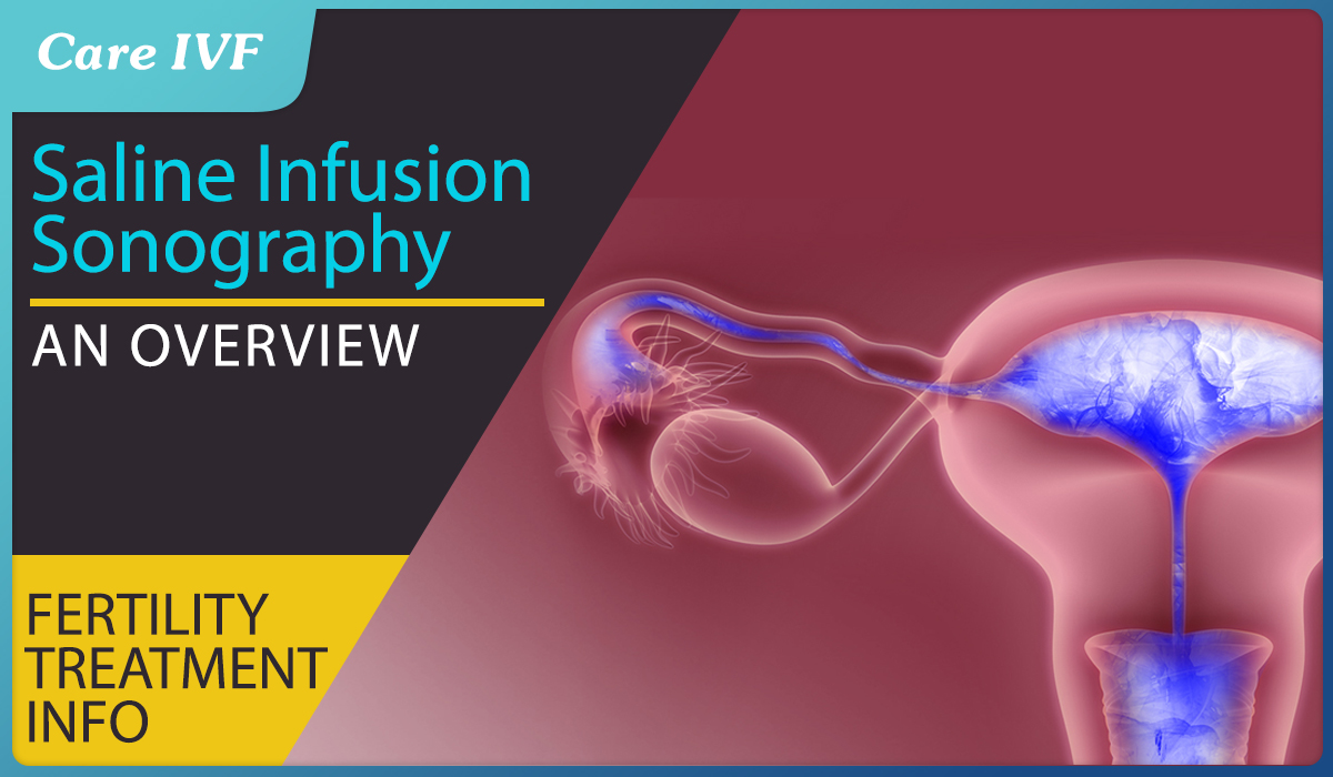
Imaging the uterus better with Saline Infusion Sonography (SIS)
The uterus is the organ where the embryo implants and the fetus grows. After the egg is fertilized by the sperm in the fallopian tube, it develops into the embryo and, 5 to 7 days post fertilization, travels down into the uterus where it implants. Abnormalities inside the uterine cavity can lead to failure of implantation of embryo, pregnancy loss, repeated miscarriage, preterm labour, and other complications.
Evaluation of the uterine cavity is hence, an essential step in fertility evaluation as well in women with history of pregnancy loss. The uterus should be mandatorily checked for fibroids, polyps, adhesions and structural abnormalities prior to IVF. While a 2D ultrasound is a standard part of investigation an infertile couple or in women with pregnancy loss, it may not detect all cavity defects. This is where a saline infusion ultrasound (SIS) can help
What is saline infusion sonohysterography (SIS)?
Saline infusion sonohysterography (SIS) or Sonohysterogram (SHG) is a simple technique in which a small amount of saline is inserted into the uterus that allows the lining of the uterus to be clearly seen on an ultrasound scan.
How do I prepare for a SIS?
No special preparation is required. The scan is best done as your period finishes, day 5–9 of your menstrual cycle. So, it is best to arrange your appointment according to your period dates.
You will be asked to go to the toilet and empty your bladder before the scan. It is a good idea to wear comfortable clothing that gives easy access to the lower part of your body
An SIS scan cannot be carried out if you are pregnant or if you have a pelvic inflammatory disease. You must advise your referring doctor or staff where you are having the scan if you have either of these conditions.
Who does the SIS?
The examination is always carried out by a specialist doctor, usually a radiologist or an obstetrician/gynaecologist qualified to carry out ultrasound examinations. The specialist doctor will provide your referring doctor with a report of the scan.
Where is a SIS done?
The examination is carried out in a radiology department of a hospital, private radiology practice or at a specialist clinic for obstetric and gynaecological imaging. It is done in the privacy of an ultrasound room, which may be dimly lit to allow
How is SIS performed?
SIS is usually done after the menstrual period finishes. The procedure begins with an ultrasound examination using a probe placed in the vagina.
Next, a speculum is introduced and a narrow catheter is placed in the vagina, through the cervix, and into the uterine cavity. The ultrasound examination is continued while sterile saline (salt water) is put into the uterus. The saline solution fills the uterus, helping to outline the uterine walls and cavity. This shows abnormalities such as fibroids, polyps, or scar tissue inside the uterus.
Are there any after effects of a SIS?
After the scan, there is a small trickle of fluid from the vagina. This is the saline fluid that was inserted through the catheter coming out. It is commonly slightly blood stained and this may continue for 24 hours. You may wish to use a sanitary pad, but you are advised not to use tampons for the rest of the day.
Most patients feel normal after the scan with no after effects. Some patients may have some pelvic discomfort (like mild period pain), but this settles after a few minutes up to perhaps an hour or so, and is very uncommon.
A very small number of patients may have some dizziness due to the cervix being slightly irritated by the catheter. This usually passes within a few minutes and has no adverse outcome.
You will generally be well enough to drive home and resume normal activities, such as going back to work.
How long does a SIS take?
The entire procedure usually takes approximately 30 minutes. Most of this time is taken up by scanning before and after the saline is put into the uterus. The actual time taken for the saline to be put in is only 2–3 minutes.
What are the risks of SIS?
The scan is very safe. The main risk is that of infection within your uterus being introduced by the procedure. This is extremely uncommon and is treated with antibiotics if it occurs.
Infection may present as pelvic pain that does not settle or you may develop an odorous vaginal discharge.
In this case, you should ring the clinic where your procedure was done and ask to speak to the radiologist who carried out the procedure or see your general practitioner explaining you had the procedure and describing your symptoms. Antibiotics may then be prescribed.
What are the Benefits of a SIS?
Inserting the saline fluid into the uterus allows very clear ultrasound images to be taken of the lining of the uterus, and any uterine abnormalities, such as thickening of the endometrium or polyps, can be easily seen. This will help to guide the discussion between you and your doctor about any further investigation or treatment that may be needed.
The images on the ultrasound machine to be clearly seen by the person carrying out the scan.
What are the advantages of a SIS?
Due to the enlargement of the uterus by infusion of saline, there is enhancement in the quality of the images which helps in detecting Uterine abnormalities better. Polyps in the endometrium, fibroids that protrude into the cavity of the uterus and scarring inside the uterus are all better visualized by SIS in comparison to a 2d ultrasound. When combined with 3d ultrasound, SIS has a high detection rate for structural defects inside the uterus. It is a simple, minimally invasive test that avoids the use of radiation.
What are the risks and complications?
SIS is a very safe procedure and usually is performed without incident. Serious complications are rare. The most common serious complication with SIS is pelvic infection. However, this occurs less than 1% of the time and usually occurs when a woman also has a block or infection of the fallopian tubes.
SIS may also cause cramping, spotting, and vaginal discharge. Some women have cramping for several hours after the procedure. It is often recommended to take a medication such as ibuprofen before this test. Some doctors may also prescribe stronger pain medication and/or antibiotics before the procedure. You should call your doctor if you experience pain or fever in the 1–2 days after the SIS.
Article Tags
About the author
Leave a Comment