
Assessing Tubal Damage through Tubal Patency Tests
The lifetime incidence of infertility is 17% (1 in 6 couple). Tubal and peritoneal factor accounts 35% of infertility among couples and tubal factors alone cause 20%
Fallopian tube’s structure:
The 2 uterine tubes 10-12 cm in lengthy, lies each side of the uterus in the medial ¾th of the upper margin of broad ligament. Each tube consists of 2 opening and 4 parts.
Ostium-
- Uterine ostium is 1 mm in diameter and communicate with the intramural part of the tube with the lateral angle of the uterus.
- Pelvic /abdominal ostium at the lateral end of the tube at the bottom of infundibulum where it pierces the posterior layer of broad ligament, in its margin projects finger like projections known as fimbria.
- Parts
From medial to lateral tube consists of
Intramural-1cm long and transverse musculature of uterus at the junction of undus and body, narrowest part.
Isthmus-rounded cord like, length is 3 cm.
Ampulla –thin walled, dilated, tortuous 5 cm in length, fertilization takes place here, then it takes 72-96 hours by ciliary movements and peristalsis of tube to reach uterus.
Infundibulum-1 cm long.it presents pelvic ostium with fimbria.
- Structure
- Outside inward serous, muscular, and mucous coat. muscular coat consists of ciliated, secretory and intracalary.
Role in maintenance of fertility:
Tubal fimbria guide the ovulated egg into the tube, unidirectional beating of the tubal cilia and peristaltic contraction of the muscular tubal wall transport the egg and then the embryo.
Secretions from the tubal epithelium nourish the embryo.

Causes of tubal factor infertility:
Tubal factor infertility includes an array of disorder affecting 1 or more of the above components. Even in the presence of a patent tube damage to the inner musculature may result in severe impairment of tubal function. It is the only preventable cause of infertility.
- PID-major cause of tubal factor infertility. The likelihood of developing infertility related to number &episode of PID, severity of disease, length of time before treatment and the age of woman.
- Sexually transmitted disease;
- Chlamydia-approximately20% of woman affected by lower genital tract infection of which 4% develop chronic pelvic pain,3% infertility.
- Neisseria gonorrhea also causes tubal damage. STD s cause salpingitis and thus cause infertility.
- severe endometriosis major abdominal/pelvic surgery (proctocolectomy, severe deep infiltrating endometriosis surgery)
- Past history of ectopic pregnancy
- genital tuberculosis
TYPES OF TUBAL BLOCKS:
The tube can get blocked in multiple sites, which can be classified as proximal, mid-tubal and distal tubal blockage.
CAUSES:
Proximal tubal blockage-
- Intratubal mucosal debris
- Cornual polyps
- Salpingitis isthmic nodosa
- Corneal spasm (pseudo blockage)
Distal tubal blockage:
- Infection caused by sexually transmitted disease
- Pelvic tuberculosis and tubercular salpingitis
- Peritubal adhesion & tubal damage from endometriosis & previous pelvic surgery.
Evaluation of tubal factor infertility:
There are a plethora of tests for tubal factor infertility but no test is idea, all procedures consists of advantages and disadvantages. First test which was done by Rubin in1920 was tubal insufflations tests which is not used anymore. The different tests are
- Hysterosalpingography
- Hysteroslpingo-contrast-sonography
- Falloposcopy
- Salpingoscopy
- Laparoscopy and chromopartubation test
- In this discussion we will be focusing on hysterosalpingography and HyCosy which are the most commonly used daycare procedure and most widely used as a 1st step of evaluation of tubal factor infertility.
HYSTEROSALPINGOGRAPHY:
It is a radiographic imaging technique where a radio opaque contrast material is injected through the cervical canal to evaluate the endocervical canal, endometrial cavity & fallopian tube lumina.it can detect tubal obstruction. It is an excellent predictor of tubal infertility, sensitivity65%, specificity -83%
Timing - an average of 10-15 minute in which 1-2-minute time is taken for fliuoroscopic view.
Average radiation exposure to ovary - 1-2 Rads.
When to do - between day 6- day 10 of menstrual cycle because
- Chance of infection is less
- Thin endometrium lead the interpretation better
- Possibility of being pregnant is almost nil.
- Done after cessation of menstruation as during men’s intravasation can occur due to increased vascularity.
Preparation before HSG
- Cramping pain is very common, give NSAID 30 minute before the procedure to reduce the pain.
- Prophylactic antibiotic should be given to women if Chlamydia screening not done.(NICE).Azithromycin 1gm orally or doxycycline 100 mg twice daily for 7 days given.
Dye used:
Aquous based dye | Oil based dye |
No sharp outline | Very sharp outline |
No embolism | Oil embolism |
Not common | Very common |
no | May occur |
The use of oil-based media confer a therapeutic effect compared to water soluble media in term of live birth rate post procedure. But water based media preferred due to improved image quality and safety.
How many views - 3 views
1st before injecting contrast
2nd-after injecting 3-4 ml contrast uterine contour &filling defect
3rd to see the spill of contrast from the tubes.
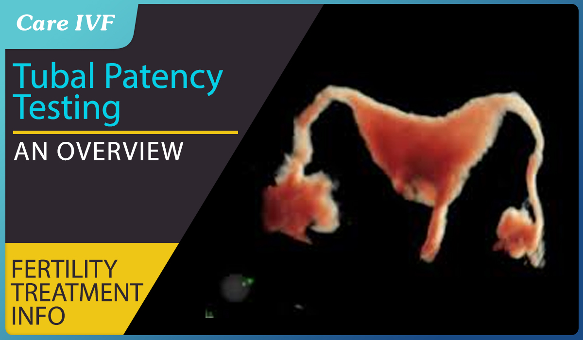
Appearance: normal endometrial cavity is triangular/ T shaped & fallopian tubes are delineated well & can be seen up to fimbrial end with spillage of dye from both tubes
normal HSG showing normal uterine contour with bilateral spillage from both the fimbrial end.
acute pelvic infection, pregnancy, adnexal mass, suspected tuberculosis, hypersensitivity to the iodine etc.
Complication: pelvic infection, vasovagal reaction, uterine perforation, allergic reaction to the contrast, intravasation of contrast (1-2%), granuloma formation with oil
based contrast etc.
Condition diagnosed by HSG
TUBES
- Tubal Blocks

By HSG site of the blocks can be determined, bi-lateral cornual block sometimes be the pseudo block due to immediate tubal spasm after injecting dye.It can be alleviated by giving sedatives 30 minutes before the procedure.
- Hydrosalpinx

A hydrosalpinx is a collection of watery fluid in the fallopian tube. with the inflammation get subsides fimbrial end get sealed but the tubal epithelium secretes the natural secretions, become distended and form hydrosalpinx. It has a very negative impact on implantation. Salpingectomy before IVF increases the live birth rate 2 times and hence recommended.
- Tuberculosis: HSG not done to detect tuberculosis, but in a n evaluation of infertility by HSG it may be detected incidentally.
Endometrial finding-synechia, irregular contour
Tubes-ragged appearance, multiple constriction or breaded appearance, tobacco pouch appearance, calcified tubes etc.

UTERUS
Acutely anteverted/retroverted
Congenital abnormalities (infantile / unicornuate / bicornuate & septate uterus)
Any intrauterine pathology (submucous fibroid, synechia)
CERVIX
Cervical incompetence
Cervical polyp/fibroid
TAKE HOME MESSAGE-
- HSG is cheap and widely available
- Largest evidence base to rule out unilateral or bilateral tubal block
- Low sensitivity &high specificity of 87%. A positive test correctly identifies blocked fallopian tube in 53% of cases whereas a negative test correctly identifies patent fallopian tube in 87% of cases.
- Limitation include-failed catheterization, false positive due to tubal spasm or debris.
HYSTEROSALPINGO CONTRAST SONOGRAPHY
- It is a TVS technique in which a water soluble contrast medium is injected into the uterine cavity using a 5F/7F catheter. The test is performed on an outpatient setting with the woman in a semi lithotomy position allowing easier access to cervical catheterization.

- Preparation for HyCoSy-
Do a urine for preg test to exclude pregnancy
Best time to do it day8-10 of menstrual cycle.
Take 2 tablet dulcolux and festal on the night prior to the procedure to
Prevent gas shadow.
To minimize pain give droteverine & diclofenac I.M injection 45 minute
Before procedure
Post procedure give doxycycline antibiotic coverage
Procedure

Do a TVS scan prior to procedure.
Insert speculum. The catheter inserted in the cx up to internal os & inflate
The balloon of catheter.
Again, insert the TVS probe, then insert the HyCoSy contrast material through
The catheter. As the contrast flows it inflate the uterine cavity and pass through
The tubes and patency are examined.
- Contrast media - the best media for cavity check is normal saline but combination of lignocaine gel, ExEm gel and purified water gives more accurate observation.
- Advantages over HSG
- Avoid radiation exposure.
- Simultaneous assessment of endometrial cavity along with tube
- More sensitive and specific than HSG.
- Results are available instantly, no need to wait for the x ray plate like HSG.
- Less painful than HSG (pain score is 1.5 which is much less than HSG 4.7)
- Can avoid the tubal spasm caused by HSG. Less error in result.
- No risk of contrast reaction or anaphylaxis
- Risks
- Persistent foul smell discharge
- Lower abdominal pain
- Unexplained fever
Article Tags
About the author
Leave a Comment